Which Plexus Does the Ulnar Nerve Arise From: Sacral, Cervical, Lumbar, or Brachial?
The ulnar nerve is a crucial component of the nervous system, responsible for sensory and motor functions in the upper limb. Understanding the origin of the ulnar nerve is essential in diagnosing and treating conditions that affect its function. In this article, we will explore the various nervous plexuses and determine which plexus the ulnar nerve arises from.
Understanding the Ulnar Nerve
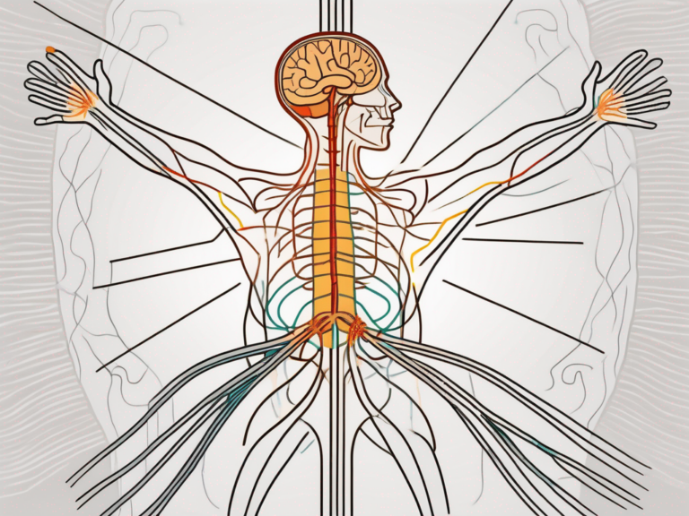
The ulnar nerve is one of the major nerves in the upper limb and plays a vital role in hand and forearm function. It originates from the brachial plexus, a complex network of nerves that arise from the cervical and upper thoracic spinal roots. The brachial plexus gives rise to several nerves that innervate different regions of the upper limb, including the ulnar nerve.
The ulnar nerve is a fascinating structure that deserves a closer look. Let’s dive into the anatomy of this nerve and explore its intricate pathway through the upper limb.
Anatomy of the Ulnar Nerve
The ulnar nerve begins its journey in the brachial plexus, specifically from the C8 and T1 spinal nerve roots. These roots unite to form the lower trunk of the brachial plexus, which then gives rise to the medial cord. The ulnar nerve arises from the medial cord, along with other branches that innervate different muscles and areas of the arm and hand.
As the ulnar nerve emerges from the brachial plexus, it embarks on a remarkable expedition through the upper limb. Its path takes it through various anatomical landmarks, each with its own significance.
After its origin, the ulnar nerve travels down the arm, passing through the axilla and medial to the brachial artery. This journey is not without its challenges, as the nerve must navigate through a crowded space filled with other structures. It is a testament to the ulnar nerve’s resilience and adaptability.
As the ulnar nerve continues its descent, it encounters a well-known landmark – the medial epicondyle of the humerus, commonly known as the funny bone. This bony prominence serves as a reminder of the ulnar nerve’s close proximity to the surface, making it susceptible to injury when struck.
Once in the forearm, the ulnar nerve embarks on the final leg of its journey. It dives deep between the two heads of the flexor carpi ulnaris muscle, a move that requires precision and coordination. This maneuver ensures that the ulnar nerve reaches its intended destination – the hand.
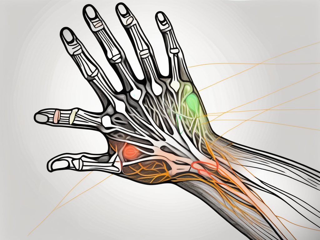
Upon reaching the hand, the ulnar nerve branches out, supplying motor fibers to several intrinsic hand muscles. These muscles are responsible for delicate finger movements and fine motor skills. Additionally, the ulnar nerve provides sensory innervation to specific areas of the hand, allowing us to perceive touch, temperature, and other sensory stimuli.
Function of the Ulnar Nerve
The ulnar nerve is a multitasker, fulfilling both motor and sensory functions in the ulnar-innervated muscles and regions of the hand. Its motor branches control the flexor carpi ulnaris, flexor digitorum profundus, and most of the intrinsic hand muscles that govern finger movements. Without the ulnar nerve, our ability to perform intricate hand movements would be severely compromised.
In terms of sensory function, the ulnar nerve provides sensation to the ulnar half of the fourth digit and the entire fifth digit, as well as the corresponding regions on the palm and dorsal surfaces of these fingers. This sensory input allows us to perceive the world around us and interact with our environment in a meaningful way.
The ulnar nerve is truly a remarkable structure, intricately woven into the fabric of our upper limb. Its role in hand and forearm function cannot be overstated, and its anatomy and function continue to captivate researchers and medical professionals alike.
Overview of Nervous Plexuses
Nervous plexuses are intricate networks of nerves that serve different regions of the body. They are formed by the branching and merging of multiple spinal nerves and allow for efficient distribution of nerve fibers to various organs and tissues.
These complex networks of nerves play a crucial role in maintaining the proper functioning of the body. By interconnecting and intermingling, nervous plexuses ensure that the communication between the central nervous system and the peripheral nerves is seamless and efficient.
Let’s delve deeper into the fascinating world of nervous plexuses and explore their structure, formation, and the important role they play in the body.
What is a Nervous Plexus?
A nervous plexus is formed as the nerves originating from different spinal roots converge and intermingle, forming a mesh-like structure. This network allows for the exchange and redistribution of nerve fibers, ensuring proper innervation of specific areas of the body.
Imagine a complex web of nerves, intricately woven together, like a masterpiece of nature’s design. Nervous plexuses are like the nerve highways of the body, connecting different regions and facilitating the flow of vital information.
These plexuses are formed by the merging and branching of multiple spinal nerves, creating a network that is greater than the sum of its parts. This intricate intermingling ensures that no area of the body is left without proper nerve supply, allowing for precise control and coordination.
Role of Nervous Plexuses in the Body
Nervous plexuses play a vital role in maintaining the functional integrity of various body regions. They enable efficient communication between the central nervous system and the peripheral nerves, ensuring the transmission of sensory and motor signals to and from specific areas.
Think of nervous plexuses as the messengers of the body, relaying important signals between different parts. They are responsible for transmitting sensory information, such as touch, temperature, and pain, from the body to the brain, allowing us to perceive and respond to our environment.
Additionally, these intricate networks facilitate the transmission of motor signals from the brain to the muscles, enabling us to move and perform complex actions. Without nervous plexuses, our movements would be uncoordinated and our senses dulled.
Furthermore, nervous plexuses are involved in the regulation of various bodily functions, such as digestion, respiration, and circulation. They ensure that the organs and tissues receive the appropriate nerve supply, allowing for optimal functioning.
Overall, nervous plexuses are essential for maintaining the harmony and balance within the body. They are the intricate wiring system that allows us to experience the world, move with precision, and function as complex beings.
The Sacral Plexus
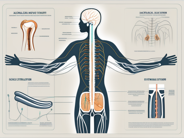
The sacral plexus is a crucial nervous plexus located in the pelvis, specifically in the posterior region. It arises from the ventral branches of the lower lumbar and upper sacral spinal nerves. The sacral plexus innervates the lower limb, buttocks, and perineum, but does not contribute to the innervation of the ulnar nerve.
Anatomy and Function of the Sacral Plexus
The sacral plexus is a complex network of nerves that plays a vital role in the functioning of the lower body. It is responsible for providing both motor and sensory function to various regions. The nerves originating from the sacral plexus innervate the gluteal muscles, hamstring muscles, and the muscles of the leg and foot. This intricate web of nerves allows for the coordination and control of movements such as walking, standing, and maintaining balance.
One of the key components of the sacral plexus is the sciatic nerve, which is the longest and thickest nerve in the human body. The sciatic nerve originates from the sacral plexus and extends down the back of the thigh, branching out to provide motor and sensory innervation to the muscles and skin of the leg and foot. It is responsible for transmitting signals that allow us to perform essential activities such as walking, running, and even simple tasks like flexing our toes.
In addition to the sciatic nerve, the sacral plexus also gives rise to other important nerves, including the pudendal nerve. The pudendal nerve is responsible for providing sensation to the perineum, which is the area between the anus and the external genitalia. It plays a crucial role in sexual function and is involved in the control of bowel and bladder movements.
Does the Ulnar Nerve Arise from the Sacral Plexus?
No, the ulnar nerve does not arise from the sacral plexus. The ulnar nerve has a different origin and is not associated with the sacral plexus. Instead, the ulnar nerve arises from the brachial plexus, which is located higher in the body.
The brachial plexus is a network of nerves that originates from the ventral rami of the lower cervical and upper thoracic spinal nerves. It provides innervation to the upper limb, including the muscles of the forearm and hand. The ulnar nerve, a major branch of the brachial plexus, travels down the arm and supplies sensation to the little finger and half of the ring finger, as well as motor function to certain muscles in the hand.
It is important to understand the distinct origins of nerves like the ulnar nerve and the sacral plexus, as they have different functions and play unique roles in the overall functioning of the human body.
The Cervical Plexus
The cervical plexus is another important nervous plexus located in the neck region. It is composed of the ventral branches of the upper cervical spinal nerves, specifically C1 to C4. The cervical plexus innervates various muscles in the neck and plays a crucial role in neck movement.
The cervical plexus, with its intricate network of nerves, is a fascinating structure that contributes to the complex functioning of the neck. It is responsible for transmitting signals from the brain to the muscles, allowing for coordinated movement and stability. Without the cervical plexus, simple actions like turning our heads or tilting our necks would be impossible.
Anatomy and Function of the Cervical Plexus
The cervical plexus supplies motor fibers to the muscles responsible for neck flexion, extension, and rotation. These muscles work together seamlessly, allowing us to perform a wide range of movements with ease. Additionally, the cervical plexus provides sensory innervation to specific areas of the neck, including the skin over the ear and the back of the head.
Imagine the intricate web of nerves within the cervical plexus, intricately connected to each other and branching out to different regions of the neck. These nerves not only enable us to move our necks but also play a vital role in our ability to feel sensations in this area. The sensory innervation provided by the cervical plexus allows us to perceive touch, temperature, and pain, ensuring our safety and well-being.
Does the Ulnar Nerve Arise from the Cervical Plexus?
No, the ulnar nerve does not arise from the cervical plexus. The ulnar nerve originates from a different part of the brachial plexus. While the cervical plexus focuses on the neck region, the brachial plexus extends further down the arm, providing innervation to the upper limb.
The brachial plexus, like the cervical plexus, is a complex network of nerves that ensures the proper functioning of the arm and hand. It is responsible for transmitting signals from the spinal cord to the muscles and skin of the upper limb, allowing us to perform intricate movements and perceive sensations.
Understanding the intricate connections and divisions within the nervous system is crucial in comprehending the complexity of our bodies. While the cervical plexus and brachial plexus are distinct entities, they work together harmoniously to ensure the proper functioning of our necks, arms, and hands.
The Lumbar Plexus
The lumbar plexus is a complex network of nerves located in the lower abdominal region. It is formed by the ventral branches of the upper lumbar spinal nerves, specifically L1-L4. This intricate web of nerves plays a crucial role in innervating various muscles and providing sensory information to specific regions of the body.
One of the primary functions of the lumbar plexus is to supply motor fibers to the muscles of the anterior and medial thigh. These muscles include the powerful hip flexors, such as the iliopsoas and the quadriceps femoris. The iliopsoas muscle, consisting of the psoas major and iliacus muscles, plays a vital role in flexing the hip joint and is essential for activities such as walking, running, and climbing stairs. The quadriceps femoris, a group of four muscles located on the front of the thigh, is responsible for extending the leg at the knee joint.
In addition to its motor functions, the lumbar plexus also provides sensory innervation to specific areas of the body. The skin overlying the anterior and medial thigh, the lower abdomen, and the inguinal region all receive sensory input from the lumbar plexus. This means that any sensations, such as touch, pressure, or temperature, experienced in these regions are transmitted through the lumbar plexus to the brain for interpretation.
Anatomy and Function of the Lumbar Plexus
The lumbar plexus is a complex network of nerves that arises from the ventral branches of the upper lumbar spinal nerves. It is situated deep within the lower abdominal region, nestled among the muscles and organs. The plexus consists of multiple nerve branches that extend and intertwine, forming a intricate web-like structure.
As mentioned earlier, one of the key functions of the lumbar plexus is to supply motor fibers to the hip flexor muscles. The iliopsoas muscle, which is innervated by the lumbar plexus, is responsible for flexing the hip joint. This action is crucial for various activities, including walking, running, and even sitting down. The quadriceps femoris, another muscle group innervated by the lumbar plexus, is responsible for extending the leg at the knee joint, allowing for movements such as kicking or jumping.
In terms of sensory innervation, the lumbar plexus plays a vital role in transmitting sensory information from specific regions of the body to the brain. The skin overlying the anterior and medial thigh, for example, receives sensory input from the lumbar plexus. This means that any sensations, such as touch, pressure, or temperature, experienced in this area are detected by the sensory nerves of the lumbar plexus and relayed to the brain for interpretation.
Does the Ulnar Nerve Arise from the Lumbar Plexus?
No, the ulnar nerve does not arise from the lumbar plexus. It is important to differentiate between the lumbar plexus and other nerve networks, such as the brachial plexus, where the ulnar nerve does originate. The brachial plexus is a network of nerves located in the upper limb region, specifically in the shoulder and arm. It is responsible for innervating the muscles and providing sensory information to the upper limb.
The ulnar nerve, which is a branch of the brachial plexus, supplies motor fibers to various muscles in the forearm and hand. It also provides sensory innervation to specific regions of the hand, including the little finger and half of the ring finger. This nerve plays a crucial role in fine motor control and sensation in the hand, allowing for precise movements and tactile perception.
While the lumbar plexus and the ulnar nerve are both important components of the nervous system, they arise from different regions of the spinal cord and serve distinct functions in the body.
The Brachial Plexus
The brachial plexus is a complex network of nerves located in the neck and axilla. It supplies motor and sensory function to the upper limb.
Anatomy and Function of the Brachial Plexus
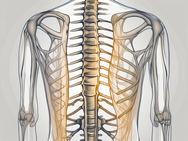
The brachial plexus consists of roots, trunks, divisions, cords, and branches. It originates from the ventral branches of the lower cervical and upper thoracic spinal nerves. The brachial plexus provides motor innervation to the muscles in the shoulder, arm, forearm, and hand. It also supplies sensory fibers to different regions of the upper limb.
Does the Ulnar Nerve Arise from the Brachial Plexus?
Yes, the ulnar nerve does arise from the brachial plexus. More specifically, it originates from the medial cord of the brachial plexus.
Conclusion: Origin of the Ulnar Nerve
In conclusion, the ulnar nerve arises from the brachial plexus, which is comprised of nerves originating from the cervical and upper thoracic spinal roots. It does not originate from the sacral, cervical, or lumbar plexus.
Summary of Findings
To recap:- The ulnar nerve arises from the brachial plexus. – The sacral plexus does not contribute to the innervation of the ulnar nerve.- The cervical plexus does not give rise to the ulnar nerve.- The lumbar plexus is not involved in the ulnar nerve’s origin.
Implications for Medical Practice
If a patient presents with ulnar nerve-related symptoms, understanding the nerve’s origin from the brachial plexus can aid in proper diagnosis and treatment. It is essential to consult with a medical professional who can accurately assess the condition and develop an appropriate management plan.
Note: This article is for informational purposes only and should not replace medical advice. If you have any concerns or questions regarding the ulnar nerve or any other medical condition, please consult with a healthcare professional.
